Now Offering
Cone Beam CT imaging of the head and neck.
Precision, Clarity, Confidence: Why CBCT imaging is the future of care.
We are excited to bring new and incredible technology to our practice members and to Corpus Christi. Cone Beam CT Imaging of Corpus Christi is the 1st accredited imaging center in South Texas with specific 3D imaging of the head and neck.
The Future of Upper Cervical Chiropractic
Cone Beam CT Imaging of Corpus Christi is located inside the office of Family Wellness Chiropractic Center.
What is Cone Beam Computed Tomography (CBCT)?
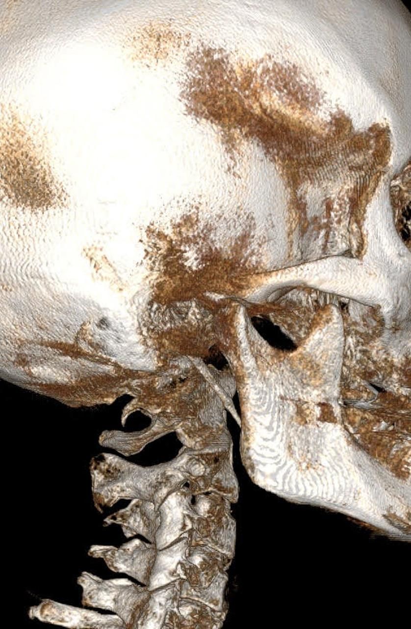
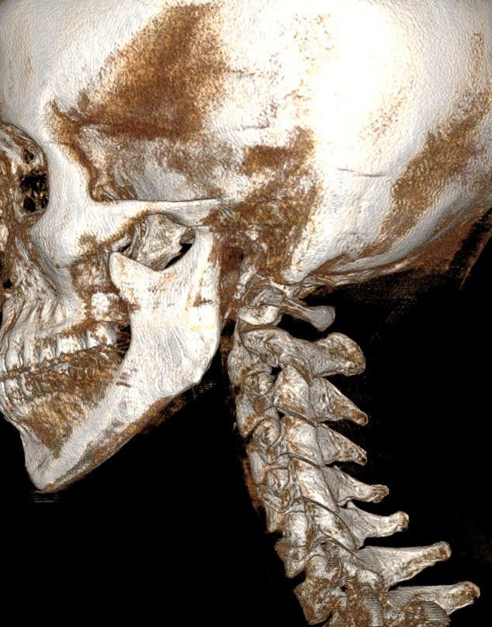
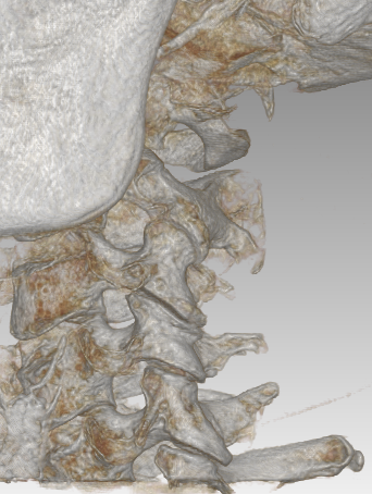
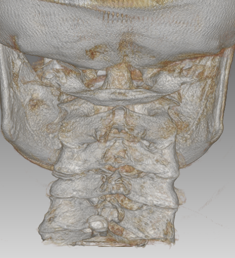
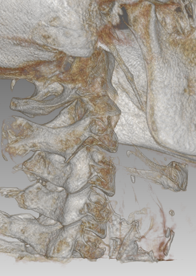
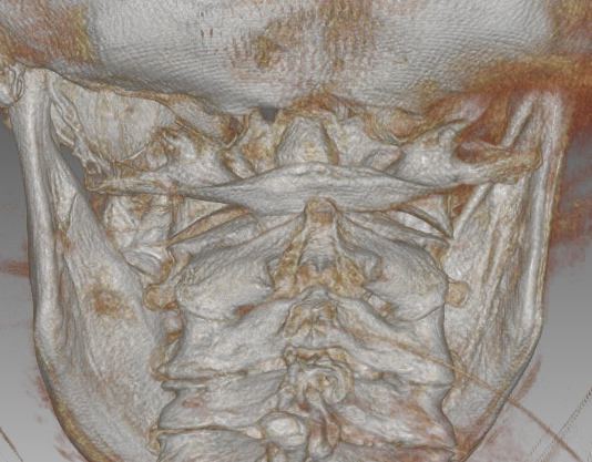
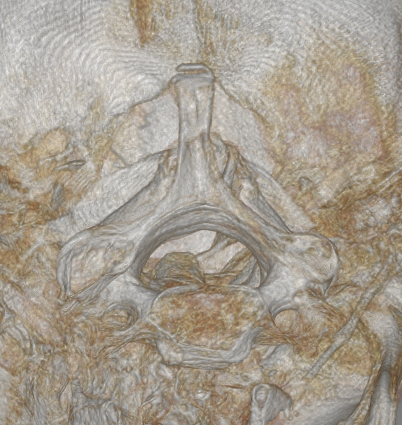
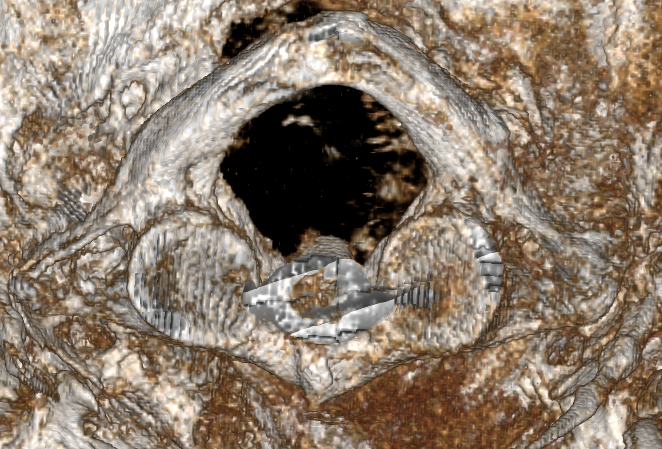
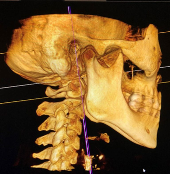
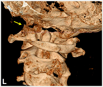
Cone beam computed tomography (CBCT) is a radiographic imaging method that allows accurate, three-dimensional imaging of the bony structures of the head (cranium), and neck (cervical spine).
Next Level Imaging
We now utilize the most state-of-the-art 3D imaging for our patients. This allows us to visualize and measure the upper neck subluxations/misalignment in a way that is completely revolutionary in the Chiropractic profession, as well as in other healthcare professions.
Cone Beam Computed Tomography (CBCT) is a specialized type of X-ray that can view the Cranio-Cervical Junction in 3 Dimensions (3D). Unlike traditional X-rays that take multiple 2-D images to attempt to view the 3-D human structure, the Cone Beam imaging can view all 3 dimensions in one scan. And these scans are incredibly fast (30 seconds), as well as extremely low dose radiation (10%) as compared to a traditional CT scan. The imaging beam is a ‘cone-like’ formation that is moved around the patient 180-360 degrees to produce the highest quality 3D images. The CBCT machine can capture hundreds and hundreds of angles in a single scan.
How It Started
Cone Beam CT Imaging of Corpus Christi was established by Scott G. Walker, DC, and is situated within his Chiropractic office, Family Wellness Chiropractic Center. Dr. Walker was introduced to this remarkable technology and the exceptional, detailed images it produces, which enabled him to seek answers for his wife, who had been enduring chronic neck pain for several years following a car accident in her teens. Despite undergoing MRIs, digital motion, X-rays, and other diagnostic testing, we were unable to identify the underlying cause of her condition. In her own research, she found a few head and neck specialists around the country using CBCT to diagnose and treat difficult conditions. We were able to find a Chiropractor in San Antonio, who has a CBCT machine, and we made the trip to have her scanned. And upon discovering CBCT, we gained a clearer understanding of the situation, which prompted us to revise our approach to her care.
With a new care plan in place, this greatly improved her condition and has helped her to function better with very few symptoms if any. This positive outcome coupled with market research, led us to recognize the limited utilization and availability of this technology in South Texas. Consequently, we made the decision to invest in this technology and offer it as a service to our current and prospective patients. Additionally, we intend to make this service accessible to our surrounding community, providing those seeking answers with a comprehensive diagnostic tool.
Don’t wait for answers – schedule your CBCT scan now!
What is CBCT used for?
This modern technology serves to increase the accuracy of the patient’s diagnosis and ultimately lead to superior outcomes while emitting less radiation. Until recently, CBCT imaging has only been used in the dental and ear nose and throat industry with its increased detail and higher performance, as that was the intended design. With more powerful units being designed recently, the Upper Cervical Chiropractic world has found this imaging to be quite beneficial for better patient diagnostics and results. The Cone Beam CT imaging is superior to traditional X-rays and can now be utilized in a chiropractic setting. It provides a three-dimensional (3-D) reconstruction of individual cervical vertebrae and their relationship to one another. It has many applications that pertain to head and neck pathologies and can be utilized by head and neck specialists including Chiropractors, Orthopedic surgeons and Neurosurgeons.
This technology will help to see things that we otherwise are not able to see with other imaging. Allowing us to improve our care plans and recommendations by being more specific in our adjustments and techniques to provide the best care possible.
What are the advantages of CBCT?
The advantages of CBCT scanning compared to most radiographic imaging techniques include the following:
➢ Can be performed with the patient in standing or sitting positions
➢ Patients can be placed in rotational or other symptomatic head and neck positions.
➢ Emits low amounts of radiation (10%) compared to conventional CT scans.
➢ Improves diagnostic capabilities which leads to better patient outcomes.
➢ Clear visualization of the Styloid bones. An elongated calcified styloid process (Eagles Syndrome) can compress the carotid sheath, especially closing off the jugular veins, internal carotid arteries leading to intracranial hypertension, as well as various cranial nerves like the Vagus nerve (brake pedal of the nervous system).
➢ Provides 3-D vs 2-D images.
➢ Can perform a full scan in about 30 seconds.
➢ Instability of the upper cervical spine and assess things like the occipito-atlanto joint (C0-C1), facial and cervical soft tissue densities, congenital (developmental) anomalies, empty sella syndrome, and other brain pathologies.
➢ Can visualize hard-to-find images of the head and neck to include being used to identify density of bone, fractures, skeletal maturation, cervical fusion assessments, cervical and temporomandibular joint instabilities, bony alignment, styloid pathologies, paranasal sinuses, sleep apnea (narrowed airway spaces), blood vessel and brain tissue (intracranial) calcifications, semicircular canal (temporal bone, structures in the inner ear and middle ear can be seen), cervical osteoarthritis, joint swelling, TMJ syndrome
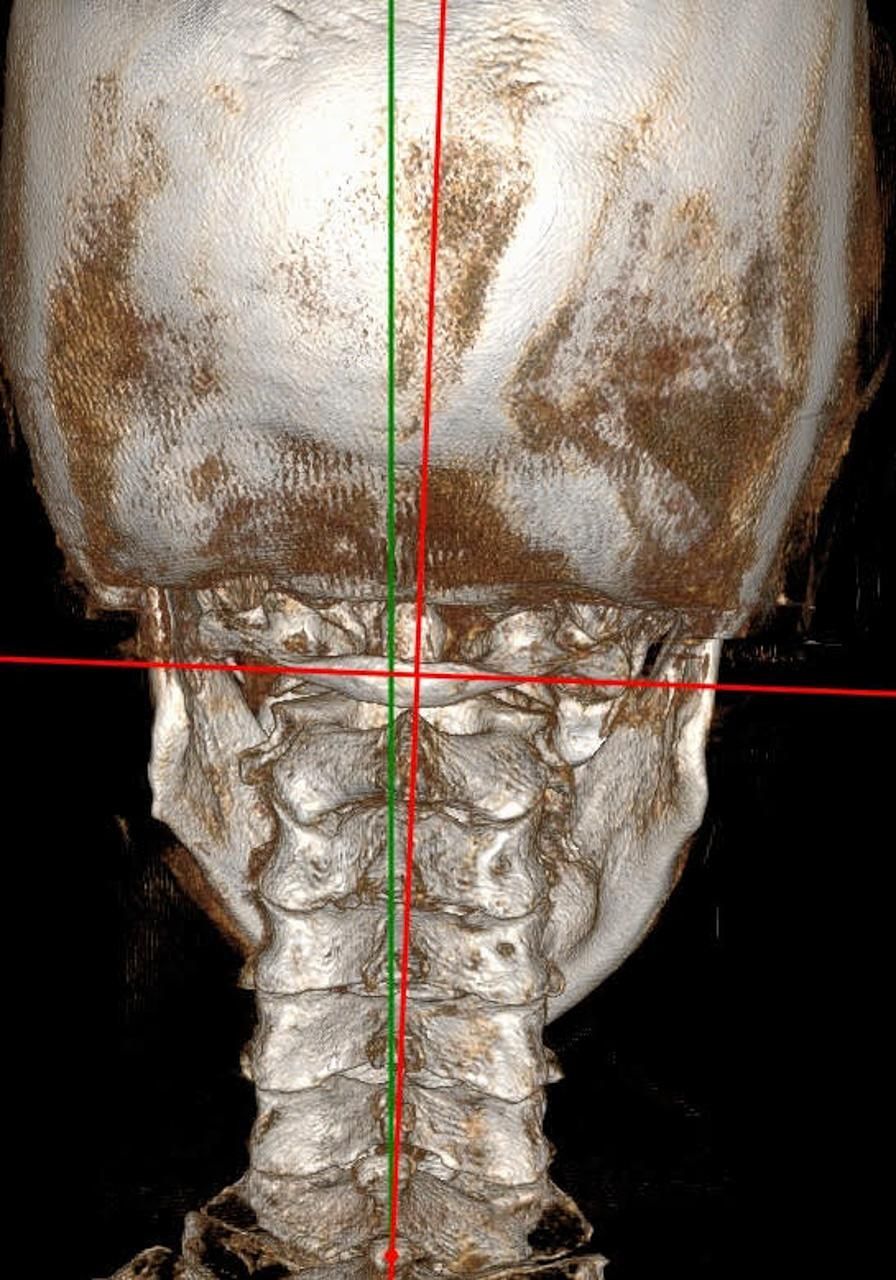
➢ The series of images then can produce projection images that can be seen in all three orthogonal planes (axial, sagittal, and coronal). The software program then allows the reconstruction of the images to give a three-dimensional image that allows an almost limitless possibility of important measurements including the following:
- Multiple measurements and angles at the craniocervical junction, including odontoid process and atlanto-dens interval. Orthospinology, Atlas Orthogonal, Blair, Pierce and other upper cervical techniques use measurements to achieve more specific upper cervical adjusting. Spinal canal volume at each vertebral segment. 3-D spatial arrangements between cervical vertebrae, facial bones, and the cranium and cervical vertebrae. Cervical vertebral bony and joint subluxations in the various planes. Neuroforaminal dimensions.
- Degree of upper and lower cervical instabilities and in what plane.
- Upper airway spaces (helpful in sleep apnea.).
- The distance between facet joints to show gapping or swelling. Atlantostyloid interval (ASI) – distance between the styloid bone and atlas.
- The displacement of the atlas in 3 dimensions. C6-atlas interval (C6-AI) – distance between the posterior C6 vertebral body and anterior arch of atlas (C1). (Shows how far forward the top of cervical vertebrae is compared to lower segments). This looks at head forward posture.
How is CBCT performed and used at Cone Beam CT Imaging of Corpus Christi?
While in the desired upright position, the scanner moves around the patient’s head, for 30 seconds. The patient is not in a claustrophobic tube or laying down.
All CBCT scans will be read by a Radiologist with detailed report included with your scan. As well as a Chiropractic analysis upon request using specialized software to measure with precision the degree of subluxation and misalignments giving your Chiropractor the ability to know exactly where and how best to adjust you.
Advanced Imaging, Better Outcomes – book your CBCT today!
Identification of the underlying cause of symptoms:
The day-to-day stressors on the human neck, especially in the upper cervical spine can involve hundreds of pounds of force each day as the average person is in forward head posture for an average of 5-6 hours per day looking at cell phones, tablets, and computer screens.
Head forward posture lifestyle effects:
Research has documented those changes in cervical spine structure, especially a breakdown of the cervical facet joints and subsequently the normal optimum cervical lordotic curve, cervical subluxations can lead to dire consequences to the human brain and body’s autonomic and central nervous systems, as all nerve tracts and fluid flow into and out of the brain through the neck.
Unfortunately, the face-down-forward-head-posture that most of us are in throughout the day puts incredible stresses on not only the ligaments of neck, leading to bone spur formation through the cervical spine. These long-term stresses as well as trauma can also cause calcification of the atlanto-occipital ligament leading to formation of a ponticulus posticus or what is known as Kimerle’s anomaly. Which along with instability can compress the vertebral artery and/or C1 nerve root.
CBCT provides a 3-D picture of the cervical spine and its relation to the cranium, especially whether atlas misalignments/subluxations or displacements are diminishing foramen magnum space. Additionally as mentioned above, ligament calcifications are not always seen clearly on other imaging. We wanted an in-house method to accurately access them and more definitively recommend solutions for patients care. This information is used by our team to know how to best approach your care.
Fees
Your path to precision care starts here – book your CBCT scan today
Although we do not accept insurance. This specialized imaging can be supported by your FSA, or HSA cards. It is possible that you may be eligible for partial re-imbursement from your insurance provider (this is between you and your insurance company and your benefits with them). We do not and will not submit billing to any insurance company but may provide a superbill listing the services for diagnostic imaging.
